If you dream it, let it bloom


Begin Your Journey
100% Focused on You
Fertility
Diagnosis
Evaluate your fertility with a complete and personalised assessment. Discover your options for becoming a parent and plan your treatment alongside the specialists at FIV Valencia.
Book an Appointment Learn MoreFertility
Treatments
Our team of specialists has extensive experience in diagnosing and treating infertility. We will study the best options for you and, with a multidisciplinary approach, find the ideal solution to help you fulfil your dream of starting a family.
Book an Appointment Learn MoreFertility
Preservation
If you want to postpone parenthood and use your own eggs or sperm in the future, ask about vitrification. At FIV Valencia, we make it simple for you.
Book an Appointment Learn More
Your First Visit
Individualised Diagnostic Assessment
The Individualised Fertility Diagnostic Study (EDI) at FIV Valencia includes a review of your medical history, a 3D ultrasound, an anti-Müllerian hormone analysis, and, if necessary, a sperm analysis or seminogram. In just 90 minutes, you’ll have your test results and, with the guidance of a specialist in Assisted Reproduction, we’ll help you design a tailored treatment plan.


Your Family, Your Way
At FIV Valencia, we care for your fertility by offering personalised options tailored to your needs. With comprehensive care, advanced technology, and an individualised approach, we help you plan your journey with the best professionals in Assisted Reproduction. At FIV Valencia, we welcome all types of families:
Motherhood Without a Partner
Women choosing to pursue motherhood on their own can explore various Assisted Reproduction treatments using donor sperm. At FIV Valencia, we’ll propose the most suitable solutions for you.
Learn moreHeterosexual Couple
Together, we’ll determine the ideal treatment to achieve the best results. We’ll provide detailed and transparent information about the most appropriate techniques and solutions for your case.
Learn moreLesbian Couple
FIV Valencia offers a range of treatments for lesbian couples who want to have a child: Artificial Insemination, In Vitro Fertilisation with donor sperm, the ROPA Method, or Embryo Adoption.
Learn moreFertility Preservation
Plan your reproductive future by preserving your fertility. Freeze your eggs, sperm, or embryos to keep your options open for parenthood when the time is right.
Learn more
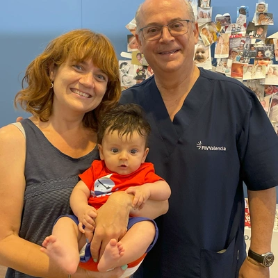
“At FIV Valencia, every case is treated with the care and dedication everyone deserves. We use effective methods that respect the health of both mother and embryo, ensuring overall well-being and a happy family with a healthy baby at home.”
Dr Miguel Dolz Medical Director, GynaecologistWhy Choose FIV Valencia?
Expertise in High-Complexity Cases
Personalised Diagnosis and Treatments
Comprehensive Care
Continuous Support
Clear and Open Communication
About us

At FIV Valencia, we believe every patient deserves a unique treatment tailored to their needs. Our expertise in Assisted Reproduction allows us to offer personalised solutions, thanks to a multidisciplinary team combining advanced technology with a warm and caring approach.
What matters most to us is being with you every step of the way. During your fertility treatment, we provide continuous support both medically and emotionally. Our commitment is to help you achieve your dream of starting a family.